Genetics Vocabulary
Genetics is an exceedingly complicated field. In order to facilitate discussions between participants and researchers, we have provided some basic genetics vocabulary.
Genome: All the genetic information a person has. Think of it like a set of encyclopedias about how to make a person.
Chromosome: A structure in cells that organizes DNA. This is like the separate volumes of an encyclopedia set.
Gene: A portion of the genome that codes for a protein. Genes are what are passed through generations creating inheritance. These are the specific chapters of each volume.
Allele: The version of a gene a person inherited. Ex: Everyone has a gene for hair color. Some people have a lighter colored allele and some have a darker colored allele. This is like the font the chapter is printed in.
DNA: The raw, most basic genetic material. Its sequence determines the structure of proteins. Think of it like the letters printed in the encyclopedia; change a letter and you can change the word.
Wild-Type: The allele found most often in a population. This is the version of the encyclopedia most people have.
Mutant: An allele found less often than the wild-type. Think of this as a typo or spelling mistake in our encyclopedia that most people don’t have.
Knockout: Complete removal of a gene to study its function.
Genotype: The alleles in a person’s genome.
Phenotype: The traits a person exhibits due to the alleles of their genes.
Penetrance: The portion of people with a mutation who show signs of the disease the mutation causes. Can be complete or incomplete.
Variable Expressivity: Variation in the severity of the symptoms seen between individuals with the same genetic disorder.
Patterns of Inheritance
Autosomal Dominant: A person only has to inherit a single allele to display the phenotype.

Autosomal Recessive: A person has to inherit two copies of an allele for a trait to display the phenotype.

Research Process
Proteins are the functional units of life, responsible for the structure, function, and regulation of the body’s cells, tissues, and organs. The instructions to make all the proteins in the human body are contained within just over 20,000 genes, which amounts to about 1% (30 million base pairs) of the human genome. The remaining 99% of the genome (3 billion base pairs) is involved in regulating when and how much protein a given gene makes, the stability of the genome, and how the genome replicates itself. Genetic disorders are caused by one or more abnormalities in the genome that result in abnormalities in protein production and/or regulation, which subsequently leads to onset of disease. Conditions that are present from birth (congenital) are likely to be genetic disorders. The field of medical genetics involves researching the causes and inheritance of these hereditary disorders, as well as the diagnosis, management, and counseling of affected patients and their families.
Medical genetics research is a vast and fast-changing field. Currently, much of our effort goes towards sequencing families containing individuals affected by primary congenital glaucoma (PCG) or high myopia. By comparing these individual’s genome sequences to that present in the general population sequencing databases, we can find the rare/novel genetic changes that may account for their disease. Our research then focuses on proving those genome variants are sufficient to cause the disease.
The first steps in our research program involve the recruitment consenting participants from eye clinic practices and isolation of their DNA from blood or saliva samples. Next, we perform focused sequencing on the parts of the genome that are already known to cause the disease when mutated. After these regions have been screened and found to be normal, the family’s DNA samples will then go for whole-genome sequencing (WGS) to try to identify the novel cause of their disease. WGS allows us to analyze the entire sequence of a person’s genome, and while this technology is undeniably powerful, it does come with some inherent downsides. As the genome of every person contains approximately 3 billion base pairs, WGS generates a lot of data! Typically, there are 4 to 5 million base pairs that differ between an individual’s genome and that of the human reference sequence. These changes are called variants, and the vast majority of them are harmless with little to no biological effect. Through WGS, we now have the ability to gather much more information than ever before, but now we must sort through all of it to identify the ‘needle-in-the-haystack’ variant that is causing the disease.
Our filtering steps must reduce the number of identified variants from millions down to a handful of likely candidates that can be investigated in greater detail. To achieve this, we systematically remove variants that logically cannot be causing the disease. We remove variants that are present at a higher frequency in the general population than the disease frequency, we look for any segregation of the variant with the disease in the family under study, and we assess the likely effect of the variant on protein structure/function. Once we have a shortlist of candidate variant genes, our research team begins searching the scientific literature to gather all the known information on those genes and their function. We then prioritize pursuing those genes that are more likely to be relevant to eye disease.
Figure: Circos Plot to visualize the genomic variation present in a typical human DNA sample. The data used to make this figure was generated via whole genome sequencing (WGS). The outermost concentric ring contains chromosomal information, including chromosome number, Giemsa banding and genetic distance in centiMorgans (cMs). The second ring represents the depth to which the genome was sequenced in histogram style (blue bars show average coverage for every half-megabase region). The third ring represents the density of small insertion-deletions (indels) in scatter plot style (black dots represent the number of indels for each megabase of sequence). The fourth ring represents the density of single nucleotide polymorphisms (SNPs) in scatter plot style (green dots represent the number of SNPs for each megabase of sequence). The fifth ring represents the proportion of homozygous SNPs (orange) and heterozygous SNPs (grey) for each megabase region in histogram style. The sixth ring represents the presence of copy number variants (CNVs) – a red line shows a mean gain and a green line shows a mean loss of genomic material. The most central ring represents large structural variants (SVs) identified in exonic and splicing regions (translocations are orange, insertions are green, deletions are grey, duplications are pink and inversions are blue).
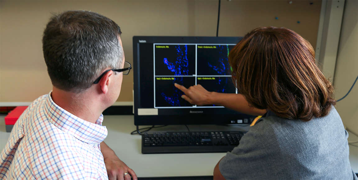
When a prioritized shortlist of candidate gene variants has been created, there are three major stages to proving which gene variant is causal for the eye disease. First, we show that the gene’s protein product is normally expressed in the same tissue that is affected by the disease. For this, we use protein-specific staining techniques on normal mammalian eye tissues. Second, we must prove that the gene variant has an effect on the function of its protein product. To this end, we express normal and variant forms of the gene in cultured cell lines and test whether or not the variant protein behaves the same way the normal protein does. Finally, in order to confirm that the gene variant is sufficient to cause the disease, we genetically engineer zebrafish or mice to harbor the same variant. This is achieved using the latest in CRISPR-Cas9 genome-editing technology. Once an animal has been engineered, we breed and age them to see whether they develop similar disease symptoms to their human counterparts. While the process of editing an animal’s genome is extremely powerful, it is also very time consuming – the generation of a single, correctly-edited, animal takes at least 8 months, with subsequent breeding and ocular assessment steps taking the investment time to well over a year.
Once a gene variant has been proven to cause the disease through animal modeling, our research focus can shift to the application of this finding. Now that mutations in a novel gene have been associated with the disease, molecular screening of this gene can be performed for other patients with the same disease. With a new gene test available, earlier molecular detection can be achieved, even while a child is in the womb.
As more and more patients are identified harboring mutations in the new gene, knowledge is accumulated as to how different mutations may give some differing disease symptoms. This allows correlations to be drawn between certain mutation types and their effect (genotype-phenotype correlations). This knowledge is important when analyzing the results of genetic testing and for subsequent informed counseling of the patient and their families.
The end goal for researching the molecular basis of genetic disease is to identify the aberrant biology behind the disease and to see which biochemical pathways may be amenable to new drug treatment options. The identification of the underlying gene defect is the first step in the road towards a new drug therapy!
High Grade Myopia
About Myopia
Myopia (nearsightedness) is one of the most common eye disorders in the world with approximately 33% of Americans being affected. The lens of the eye in myopic people does not focus light on the retina, the portion of the eye that turns light into information our brain processes. This results in objects that are farther away appearing as though you were looking through an out of focus camera with the image being blurry or fuzzy. The severity of myopia is measured in diopters with 0 being normal and anything below that being myopic. The error can come from a misshapen lens or cornea, or from an elongated eye.


High myopia is a particularly severe (-6.00 diopters or less) form of myopia in which the eye length continues increasing. This causes not only continuing vision deterioration, but the increasing length puts strain on the retina and other eye structures which results in greater risk of retinal detachments, early development of cataracts and glaucoma. This type of myopia is usually seen running in families. It can then be concluded that the disorder is inherited from the parents of the affected individual. The Young lab is currently working on genetic profiles of affected families in order to try to determine the genetic changes that result in the development of high myopia.
Our Research
The primary area of research focus in our laboratory has been the molecular genetics of high-grade myopia (refractive error of -6.00 diopters or greater). We have collected several hundred families (see example pedigree below) with high-grade myopia, and have performed genome screens to map the genes involved. Linkage mapping has identified several published candidate gene regions. We are continuing to collect families with high-grade myopia, using linkage and deep sequencing technologies to identify novel candidate genes. We believe that the findings from our research endeavor will impact the understanding of eye growth and development (myopic eyes are bigger and longer), may allow for molecular interventions to be developed to prevent the vision-threatening sequelae of high-grade myopia, and may contribute to our understanding of the genetics of common, complex quantitative trait disorders.

Recent Publications
Our research on high myopia has focused on searching for genes that, when mutated, increase susceptibility to the development of high myopia. These studies are performed towards an end goal of developing sophisticated gene tests that can alert patients to their own susceptibility and to inform care providers so they may proactively treat the patient. Our hope is that a greater understanding of the genetics of high myopia will lead to a better understanding of the pathogenesis of the disease at a molecular level. This knowledge can then be utilized in the development of molecular interventions, such as pathway-targeted drugs or other therapeutics.
Exome sequence analysis of 14 families with high myopia
This publication involved the sequencing of the protein coding portions of the genome from various families with inherited forms of high myopia. This analysis yielded many new candidate genes for consideration in further studies of high myopia.
Trio-based exome sequencing arrests de novo mutations in early-onset high myopia
This work involved sequencing family trios – families consisting of 2 non-myopic parents and a young, high myopic child – to identify spontaneously occurring (de novo) sequence changes in the affected child that had not been inherited from either parent. This study led to the discovery of BSG gene variants in many of the children. Furthermore, mice harboring mutated BSG genes were generated and found to be myopic.
The CREAM consortium (Consortium for Refractive Error And Myopia) is a large, multinational group of researchers who work to further the understanding of the basis of myopia. Our lab worked in collaboration with this consortium to identify genetic sequence variants in children that associate with refractive error and myopia, and to also assess how their environment may have altered their disease risk. This research has helped to identify candidate genes that may be relevant to early-onset myopia, while accounting for the higher environmental risks in certain individuals.
This research was also performed in conjunction with the CREAM consortium to identify portions of the genome that may be associated with myopia, but also took into account the educational level of the individuals being analyzed. Education level can be seen as a risk factor for myopia, as reading has been strongly implicated as an environmental risk factor in multiple studies.
Primary Congenital Glaucoma
About Primary Congenital Glaucoma
Primary congenital glaucoma (PCG) is a devastating early childhood eye disease accounting for 5% of childhood blindness and 18% of blind school registry. The eye maintains its shape via pressure from internal fluid (aqueous humor) that is produced by the ciliary body and circulates throughout the chambers of the eye.
In patients with glaucoma, the drain for this fluid (made up of the trabecular meshwork and Schlemm’s canal) does not function correctly. This allows the fluid to build up in the eye, increasing the pressure, which will damage the sensitive structures involved in light-sensing in the eye, such as the retina and optic nerve. If left untreated, blindness will invariably occur.

Primary congenital glaucoma (PCG) is a devastating early childhood eye disease accounting for 5% of childhood blindness and 18% of blind school registry. The eye maintains its shape via pressure from internal fluid that circulates throughout the chambers of the eye. In patients with glaucoma, the drain for this fluid does not function correctly. This allows the fluid to build up in the eye, increasing the pressure which will damage sensitive structures involved in light-sensing in the eye, such as the retina or optic nerve. If untreated, blindness invariably occurs.
 Early-Onset Glaucoma – Genetics Home
Primary Congenital Glaucoma – GeneReviews
Our Research
A newer area of investigation in our laboratory has been the molecular genetics of primary congenital glaucoma (PCG). The molecular cause of PCG is only partially understood. Mutations in CYP1B1, encoding a subunit of the cytochrome P450 complex, a protein involved in metabolism found in all kingdoms of life, are the most common cause of autosomal recessive PCG worldwide. They account for up to 87% of familial cases in some isolated populations, but only 25-27% of families in populations with heterogeneous ethnicity. Other genes with mutations known to cause PCG are LTBP2, MYOC and FOXC1 along with the TEK variant our lab found.
We are currently enrolling families with history of primary congenital glaucoma in a research study and performing genetic sequencing in order to identify novel candidate genes. We believe that findings from this research will deepen our understanding of eye development and may uncover novel mechanisms underlying glaucoma. This may lead to the development of targeted molecular interventions in the future.
We are partnering with pediatric ophthalmologists and glaucoma experts at the University of Wisconsin-Madison. Our clinical collaborators include:
Yasmin S. Bradfield, MD, FAAP
Professor
Pediatric Ophthalmology and Strabismus
Member, Childhood Glaucoma Research Network
Vice Chair of Education
Gregg A. Heatley, MD, MMM
Professor
Service Chief, Glaucoma Service
Vice Chair of Clinical Affairs
Yao Liu, MD
Assistant Professor
Glaucoma Service
Member, Childhood Glaucoma Research Network
Anna C. Momont, MD
Assistant Professor
Glaucoma Service
Melanie Schmitt, MD
Assistant Professor
Pediatric Ophthalmology and Strabismus
Director of Ophthalmic Genetics
Our clinical study is still ongoing.
Recent Publications
Our research on primary congenital glaucoma (PCG) has also utilized the latest next-generation sequencing technology (exome/whole-genome sequencing) to identify genes harboring small changes/variants that may be causal for the development of PCG. The discovery of new genes associating with a disease increases our understanding of the biology behind the condition, allows for molecular diagnostic testing and genetic counselling, and uncovers molecular pathways which might be targeted by potential therapies in the future.
Angiopoietin receptor TEK mutations underlie primary congenital glaucoma with variable expressivity
This publication outlines the work performed by members of the Young lab that led to the identification of mutations in the TEKgene as the second largest contributor to PCG. The lab was able to demonstrate that the TEK variants found in 10 families with PCG resulted in the loss of TEK protein function, impacting a signaling pathway critical for the correct formation of the drainage structures of the eye.
This publication was the culmination of research performed by a large number of labs collaborating to analyze possible areas of the genome that may be associated with various abnormalities associated with glaucoma. Specifically, the study implicated several genes as potentially influential for intraocular pressure levels and optic nerve structures. This study has provided insights into possible molecular mechanisms underlying glaucoma.
Optic disc changes are one of the hallmarks of glaucoma pathogenesis. This study, in collaboration with the International Glaucoma Genetics Consortium, studied the genomes of more than 20,000 individuals in order to search for genes that may be influencing these optic disc changes. The results contribute to an expanding body of knowledge of the molecular pathways involved in the development of glaucoma.
Intraocular pressure elevation is one of the first steps in the pathogenesis of glaucoma, eventually resulting in optic nerve damage and vision loss. This work centered on the analysis of more than 35,000 people to find regions of the genome that were inherited along with an increase in intraocular pressure. In addition, genome sites that were inherited along with glaucoma were also investigated and many sites were found to be the same as for increased intraocular pressure. This work helped to illuminate the interplay between intraocular pressure and glaucoma on a deeper, genetic level. This work was also performed in collaboration with the International Glaucoma Genetics Consortium.